


 WhatsApp


Ureteropelvic Junction (UPJ) obstruction is a mechanical or functional blockage at the critical anatomic juncture where the renal pelvis funnel empties into the upper ureter. Impairing the normal, gravity-assisted and peristaltic drainage of urine from the kidney down to the bladder, a UPJ obstruction triggers high back-pressure inside the renal collecting system. Over time, progressive hydronephrosis (kidney swelling) can lead to recurrent flank pain, pyelonephritis (kidney infections), nephrolithiasis (stone formation), and irreversible cortical thinning with loss of renal function.
While open pyeloplasty via a large flank flank incision was once the gold standard, it imposed substantial postoperative morbidity, muscle splitting, and prolonged hospital stays. Pure laparoscopy significantly reduced surgical trauma but was hindered by rigid instruments during delicate, sub-millimeter suturing in a confined retroperitoneal workspace.
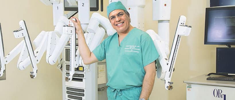
Today, Robotic-Assisted Laparoscopic Pyeloplasty represents the definitive surgical standard for repairing UPJ obstruction in both adult and pediatric populations. As Professor and Chairman of the International Robotic Institute for Prostate Cancer in South Florida, Dr. Sanjay Razdan leverages high-volume robotic mastery across more than 10,000 robotic procedures. Applying high-definition 3D visualization and wristed micro-instrumentation, Dr. Razdan performs precise, watertight reconstructive pyeloplasties that restore unobstructed renal drainage while sparing patients from debilitating incisions.
Pathophysiology and Etiology: Intrinsic vs. Extrinsic Obstruction
UPJ obstruction occurs through two primary distinct mechanisms, which directly influence the intraoperative reconstructive strategy:
[UPJ OBSTRUCTION MECHANISMS]
│
├─► 1. Intrinsic Obstruction (Aperistaltic / Fibrotic Segment)
│ └─► Congenital narrowing, collagen deposition, or mucosal scarring
│
└─► 2. Extrinsic Obstruction (Crossing Lower Pole Renal Vessels)
└─► Accessory renal artery/vein drapes over the UPJ, kinking the outflow track
- Intrinsic Stenosis: A congenital aperistaltic segment where circular smooth muscle fibers fail to develop normally, or acquired scarring from previous stone passage or instrumentation. The narrowed segment cannot propagate peristaltic waves, causing pooling of urine in the renal pelvis.
- Extrinsic Compression (Crossing Vessels): An aberrant or accessory lower pole renal artery or vein courses across the anterior aspect of the ureteropelvic junction to supply the lower kidney pole. As the renal pelvis fills, it drapes over this rigid vascular band, creating a kink that acts like a mechanical clamp on the ureter.
The Gold Standard Reconstructive Approach: Anderson-Hynes Dismembered Pyeloplasty
The most versatile and durable procedure for UPJ obstruction is the Anderson-Hynes dismembered pyeloplasty. Robotic assistance provides the visual clarity and range of motion necessary to execute this micro-reconstruction with millimeter precision:
[Robotic da Vinci Console: 3D 10x Optical Magnification]
│
▼
[Excision of Diseased Junction & Reduction of Redundant Pelvis]
│
▼
[Transposition (If Crossing Vessel Present: Anterior Mobilization)]
│
▼
[Ureteral Spatulation: Longitudinal Incision along Lateral Wall]
│
▼
[Tension-Free, Watertight Anastomosis over Double-J Internal Ureteral Stent]
- Mobilization and Anatomy Mapping: The colon is mobilized to expose the retroperitoneal space. The dilated renal pelvis and upper ureter are isolated. If an extrinsic crossing vessel is identified, the pelvic-ureteral complex is carefully mobilized so it can be transposed anterior to the blood vessels, relieving the mechanical kinking.
- Dismemberment and Excision: The diseased, aperistaltic, or fibrotic junction is excised completely. If significant chronic hydronephrosis has ballooned the renal pelvis, a reduction pyeloplasty is performed to trim redundant tissue and improve future fluid dynamics.
- Spatulation of the Healthy Ureter: The healthy, patent proximal ureter is spatulated (incised longitudinally along its lateral margin) for 1.5 to 2 centimeters. This wide spatulation ensures a broad, funnel-shaped opening to prevent future strictures.
- Endo-Wristed Running Anastomosis: Utilizing articulated instruments with 7 degrees of freedom, the spatulated ureter is anastomosed to the dependent, lowest portion of the renal pelvis using ultra-fine absorbable sutures.
- Internal Stenting: A temporary internal Double-J ureteral stent is placed across the newly constructed junction to keep it decompressed, support tension-free healing, and prevent urinary extravasation. The stent is typically removed easily in the office 4 to 6 weeks later.
Open vs. Conventional Laparoscopy vs. Robotic-Assisted Pyeloplasty
| Surgical Metric | Traditional Open Pyeloplasty | Conventional Laparoscopy | Robotic-Assisted Pyeloplasty |
| Surgical Access | 10–15 cm muscle-cutting flank incision | 3–4 tiny keyhole ports | 3–4 tiny keyhole ports (da Vinci) |
| Visualization | Limited deep pelvic/flank cavity view | 2D flat video screen | 3D High-Definition 10x Optical Zoom |
| Suturing & Articulation | Difficult through deep incision | Rigid, straight sticks (high ergonomics strain) | Wristed instrumentation (full 360° range) |
| Blood Loss & Morbidity | Moderate; rib/muscle trauma | Minimal | Negligible (< 50 mL) |
| Hospital Stay | 3 to 5 days | 1 to 2 days | Outpatient or overnight (24 hours) |
| Success Rate (Patency) | 90% to 95% | 88% to 93% | 95% to 98% durable obstruction resolution |
Diagnostic Workup and Verifying Success
Prior to surgery, precise functional and structural mapping is mandatory to confirm that the obstruction is physiologically significant:
- MAG3 Renal Diuretic Scintigraphy (Nuclear Renal Scan): Confirms delayed tracer clearance with a prolonged clearance half-time ($T_{1/2} > 20\text{ minutes}$) after administration of intravenous furosemide (Lasix), while calculating differential split renal function.
- Multiphasic CT Angiography / CT Urography: Maps out the exact vascular anatomy, confirming or ruling out the presence of crossing lower pole renal arteries and veins, while measuring the degree of hydronephrosis and stone burden.
- Postoperative Verification: Following stent removal, relief of obstruction is verified via follow-up ultrasound and a MAG3 renal scan to confirm unrestricted drainage ($T_{1/2} < 10\text{ minutes}$) and preservation of renal parenchymal function.
Conclusion
Ureteropelvic Junction obstruction does not require painful, muscle-splitting open operations or long hospital recoveries. Through robotic-assisted laparoscopic pyeloplasty, advanced reconstructive urology combines the proven durability of the Anderson-Hynes repair with the comfort and precision of robotic technology—preserving kidney function and permanently restoring urinary flow.
- Surgeon: Dr. Sanjay Razdan, MD, MCh, FRCS (Glasg)
- Academic Leadership: Professor and Chairman, International Robotic Institute for Prostate Cancer
- Official Practice Website:miamiroboticprostatectomy.com
- Main Center Location: 3650 NW 82nd Ave, Suite 501, Doral, FL 33166
- Direct Consultations: (305) 251-8650